 The CoSeC Impact Award was first launched in 2020 with three main aims: to be a means of recognising the work of researchers early in their careers who have been, or continue to be, supported by CoSeC; to be a means of raising the awareness of the communities supported through CoSeC1; and to be a means of acquiring evidence of the impact of CoSeC and the communities it supports on science, society and the economy.
This year's prizes include monetary vouchers thanks to the generous support of NAG (the Numerical Algorithms Group): £250, £100, and £75 each for the 1st, 2nd- and 3rd-places, respectively.
Congratulations to the top four applicants! Read on to learn about their winning work and future career aspirations in their own words.
1st place Ryan Warr (PhD student, University of Manchester)

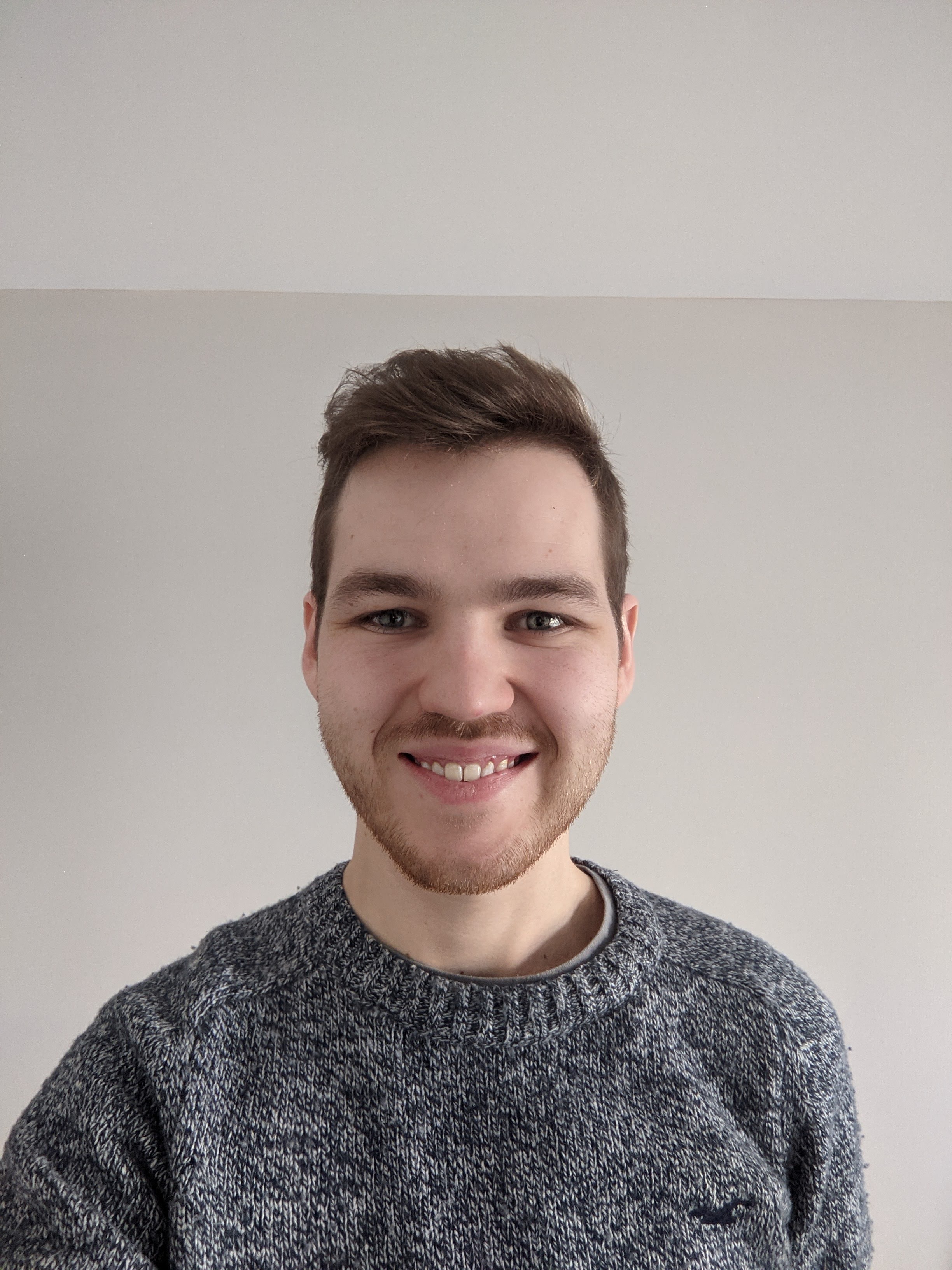 “My research concerns the advancement of hyperspectral X-ray CT for lab-based imaging, with current emphasis on improved chemical mapping of stained biological specimens through the use of novel iterative algorithms. With the support of the CCPi2, and their development of the open source Core Imaging Library (CIL) software, I have been able to demonstrate the vast improvements in elemental identification and soft tissue segmentation obtained by using spatio-spectral reconstruction algorithms created, tested and optimised using CIL. In addition to the continuous support of my research from the CCPi group, I have been able to partake in training schools that communicate the multi-modal and multi-channel reconstruction capabilities of such software. These training sessions, in collaboration with CCP SyneRBI3, not only allowed me to pass on my expertise in using these tools, but also helped improve my understanding of how they may be used for the wider imaging community.
I hope to continue my research in hyperspectral imaging in academia or industry, and help to advance it as a technique for imaging across a wide range of applications, such as geology, bioimaging and non-destructive testing. As both the imaging hardware, combined with software innovations such as CIL, continue to improve over the years, I can only see hyperspectral imaging becoming more commonplace and I hope to be there when it does!"
2nd place Dr. Antoni Wrobel (Postdoctoral Training Fellow, The Francis Crick Institute, London)

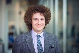 “I am a Postdoctoral Research Fellow in Steve Gamblin's lab at the Francis Crick Institute. My current work focuses on the spike glycoprotein of the coronavirus responsible for the COVID-19 pandemic, SARS-CoV-2. The spike of SARS-CoV-2 is the main protein on the surface of the virus and is responsible for its binding to the host cell. Our studies on the spike structure have provided mechanistic understanding of how the spike recognises its receptor on the surface of the host's cell and helped to understand the origin of the pandemic and importance of the newly emerging variants. This work would not have been possible without the programmes that are a part of CCP-EM4 and CCP45 packages that allowed us to determine the cryo-EM structures of the spike proteins we studied. I am currently continuing studies on the spikes of coronaviruses to better understand where the virus came from and how it became able to infect humans. In future, however, I would like to establish my own scientific programme, in which I will combine my knowledge of the viruses from the current work and that of cellular trafficking, which I gained during my PhD in David Owen's lab in Cambridge, to provide better understanding of how the viruses assemble within the cell they infected and bud from it."
Joint 3rd place Angela Harper (PhD student, University of Cambridge)

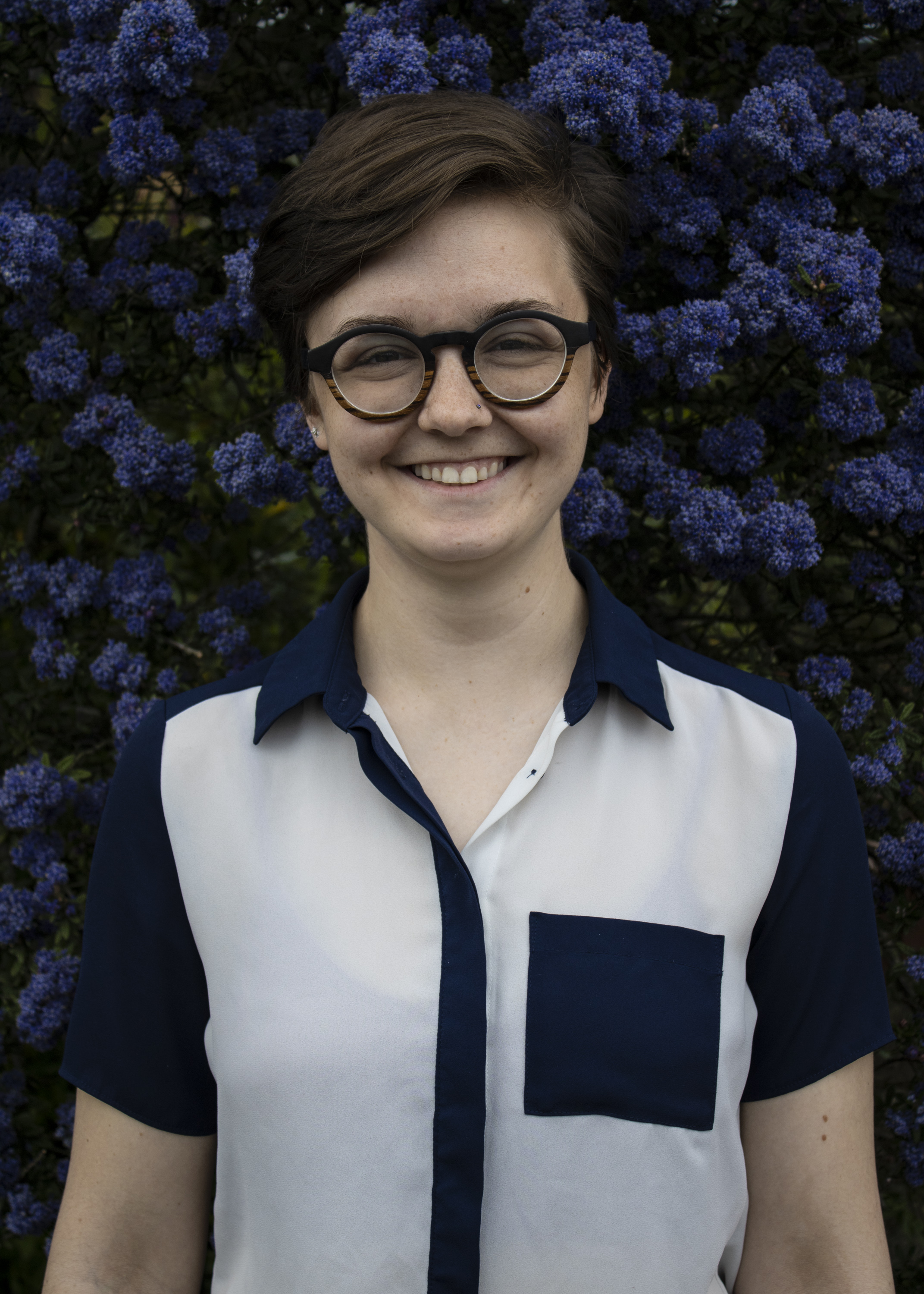 “I am currently a 3rd year PhD student in the Physics Department at the University of Cambridge. My research involves accelerating both discovery and identification of industrially relevant materials, by using ab initio methods. I have impacted each part of the discovery pipeline, by implementing workflows for prediction, characterisation, and validation using plane-wave density-functional theory (DFT) in the CASTEP code. Initially focusing my efforts on the structure prediction of crystalline materials, I have now begun work on amorphous material characterization, as many of the solid state devices at the forefront of research include amorphous materials, especially oxides. In addition to my scientific research, I have also placed a large focus during my PhD on scientific outreach, speaking at the Cavendish Inspiring Womxn series, at my college's interdisciplinary talk series, and as an alumni for my undergraduate university. Beyond my PhD, I am to continue my focus on studying industrially relevant materials, especially those with green energy applications, with the ultimate future goal of running my own lab and becoming a university professor."
Joint 3rd place Dr. Palak Wadhwa (formerly PhD student at the University of Leeds where the work for this application was carried out)

 “Computational Collaborative Project Synergistic Image Reconstruction for Biomedical Imaging (CCP SyneRBI) is focused on the development and implementation of an open-source software platform PET/MR to allow synergistic image reconstruction. My research contributed towards this collaboration by providing image reconstruction tools to reconstruct time-of-flight (TOF) PET data extracted particularly from GE SIGNA PET/MR scanner. During my research, I developed classes and utilities necessary to read the raw data outputs from the GE SIGNA PET/MR scanner and convert them into emission and data correction sinograms in Software for tomographic image reconstruction (STIR) format. These developments were then propagated to Synergistic Image reconstruction framework (SIRF) software platform. These developments also allowed to demonstrate TOF-PET image reconstruction for the GE SIGNA PET/MR data using a novel image reconstruction algorithm called TOF-kernelised expectation algorithm (TOF-KEM). My research demonstrated image reconstruction for the first time using the TOF-KEM algorithm and it was shown that the algorithm improves reconstructed PET images by substantially reducing the image noise and slightly improving image quantification. My contribution towards providing an open-source software platform to reconstruct TOF-PET data extracted from PET/MR scanner is a step towards establishing the advantages of PET/MR scanner. Since the implemented software platform during my research provides an image reconstruction framework, clinical data extracted from GE SIGNA PET/MR scanner could be reconstructed using anatomical reconstruction algorithms. These algorithms have demonstrated potential in reducing the injected dose by a factor of 10, which could benefit the vulnerable patient groups such as babies, young adults and pregnant women.
At this point in my career, my interest is to utilize the advanced image reconstruction algorithm and improve the image quality of PET images with short time frames. My research aspirations also expand towards conducting data analysis for dynamic PET data using novel radiotracers."
1These involve Collaborative Computational Projects (CCPs) and High End Computing Consortia. Read more about them
2CCPi
3CCPSyneRBI
4CCP-EM
5CCP4
